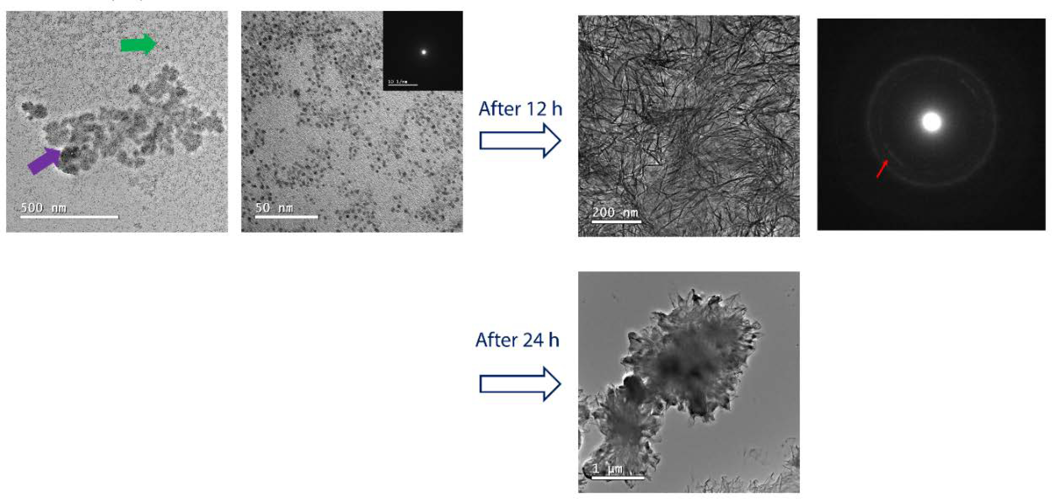
Figure 4.

Morphology of DPPS-enriched monolayers after mineralization. TEM images and their respective SAED electron diffraction patterns for the monolayers of DPPC:DPPS (8:2) molar ratio, transferred after 240 min of mineralization at 25°C. For the mixed DPPC:DPPS monolayer, the presence of nanometric complexes (~ 5 nm) indicated by the green arrow aggregates into larger structures (purple arrow). It is observed that these initially amorphous complexes crystallize after 12 h (red arrow in the SAED pattern). The formation of micrometric aggregates and a complete rupture of the transferred monolayer is observed after 24 h.
