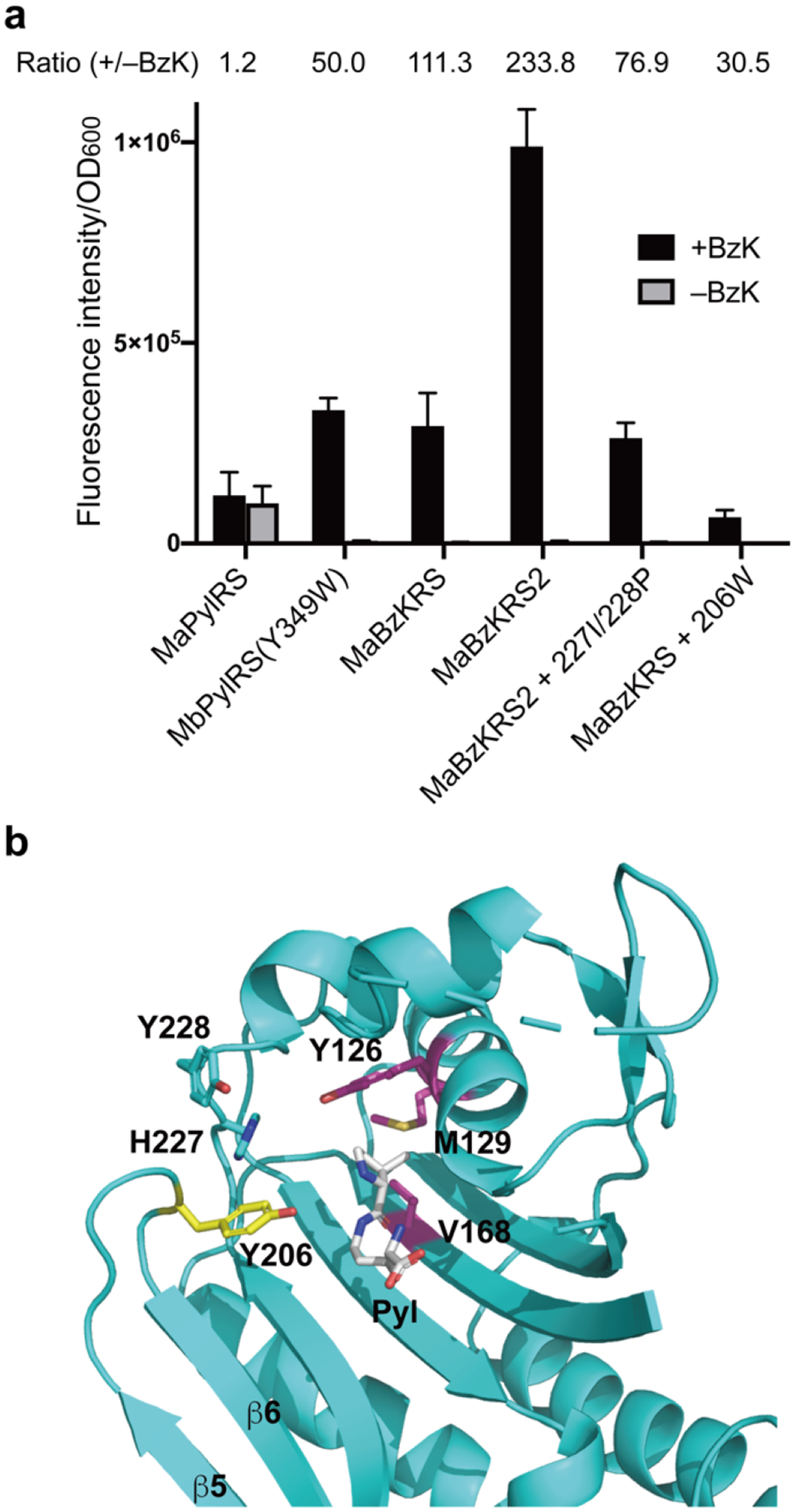
Figure 4.

Two different types of MaBzKRS for BzK incorporation. (a) Comparison of the BzK incorporation efficiency of different MaBzKRS mutants. Measured sfGFP fluorescence intensity was normalized to cell optical density. Normalized fluorescence ratio for +/− BzK was indicated for each mutant. Error bars represent s.d., n = 3 independent experiments. (b) Structure of MaPylRS showing the different locations of mutated amino acid residues in MaBzKRS (pink and cyan) and in MaBzKRS2 (yellow). To illustrate the position of the bound Pyl, M. alvus PylRS apo form (PDB 6JP2) is superimposed on M. mazei PylRSc bound with Pyl (PDB 2ZCE).
