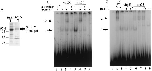
FIG. 5.
Both mouse p53 and mouse T/super T contribute to the formation of a p53-T antigen complex that can bind to DNA. (A) Silver-stained SDS-polyacrylamide gel of baculovirus-expressed T antigen purified with anti-T-antigen antibody Pab 108 from SF21 insect cells (Bacl. T) and T antigen purified from stably transformed mouse SCID cells in the absence of p53 as described in Materials and Methods (SCID T). Sizes are indicated in kilodaltons. (B) Approximately 50 ng of vhp53 or 100 ng of SCID p53 (no T antigen present; mp53) was incubated for 30 min at 30°C with 200 pg of radiolabeled probe containing the p53 binding site from RGC, 50 ng of T antigen purified from SCID cells, and 100 ng of antibody (αT) as indicated prior to electrophoresis on a 3% polyacrylamide gel. DNA-protein complexes were visualized with a PhosphoImager using Adobe Photoshop software. Arrows indicate positions of retarded complexes: 1, DNA-p53; 2, DNA-p53-T antigen. (C) Like panel B but with 100 ng of SCID p53-T antigen complex (lane 2), 100 ng of vhp53, or 100 ng of SCID p53 (no T antigen present; mp53) incubated with 50 or 100 ng of baculovirus-expressed T antigen as indicated. Arrows indicate positions of retarded complexes: 1, DNA-p53; 2, DNA-p53-T antigen.

