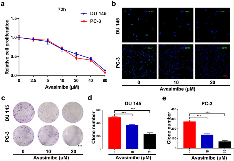
Fig. 1.
Avasimibe inhibited proliferation in PCa cells. a MTT assays were used to evaluate cell growth after treatment with various concentrations of avasimibe (0, 2.5, 5, 10, 20, 40, 80 µM) for 72 h; b immunofluorescence staining of SOAT1 to confirm the effect of avasimibe; the scale bar is 100 μm; c clonogenic survival assays were used to detect the proliferation of the avasimibe-treated PCa cell lines (PC-3, DU 145); the scale bar is 1 cm; d statistical analysis of the clone number of DU 145 cells; e statistical analysis of the clone number of PC-3 cells. Representative images are from three individual experiments. ***p < 0.001

