
Figure 3 .
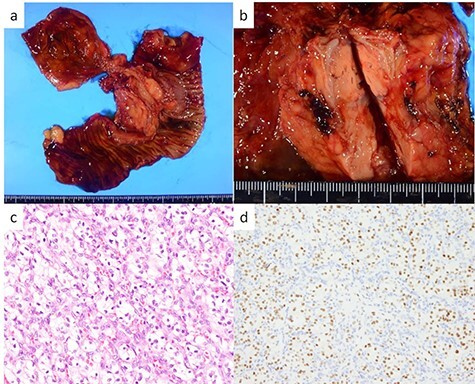
Resected specimen and pathological findings of the pancreatic head tumor; (a, b) a yellow 38-mm solid tumor in the pancreatic head protruding into the duodenum; (c) abundant nests of large clear cells (hematoxylin and eosin ×400); (d) immunohistological positivity for PAX8 (PAX stain ×200).