Figure 1.

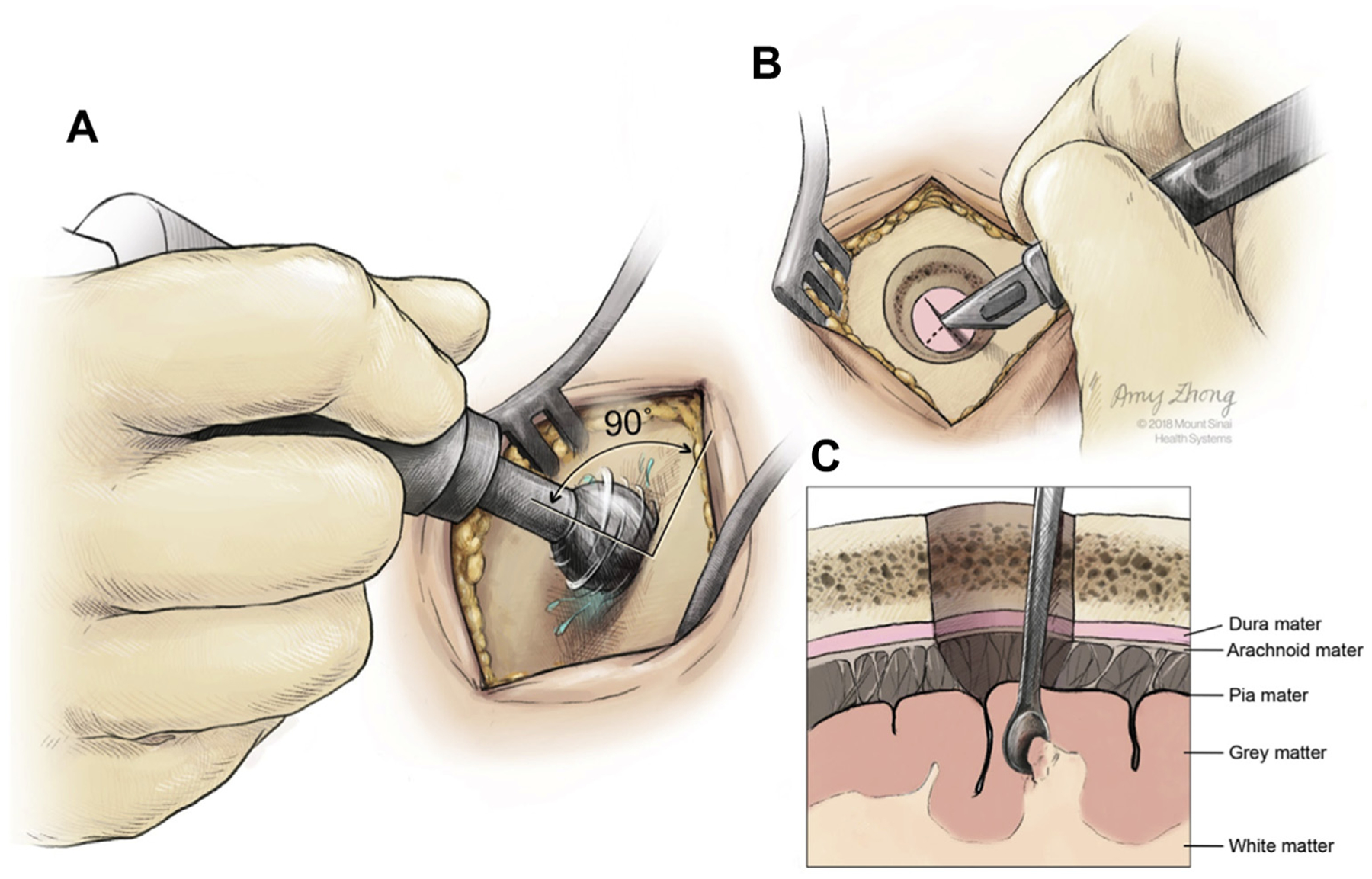
Schematic of the burr hole procedure (A), the incision of the dura (B), and the collection of a superficial biopsy at the gray/white matter junction (C). (Created by Amy Zhong at the Mount Sinai Hospital and reproduced with permission.)

Schematic of the burr hole procedure (A), the incision of the dura (B), and the collection of a superficial biopsy at the gray/white matter junction (C). (Created by Amy Zhong at the Mount Sinai Hospital and reproduced with permission.)