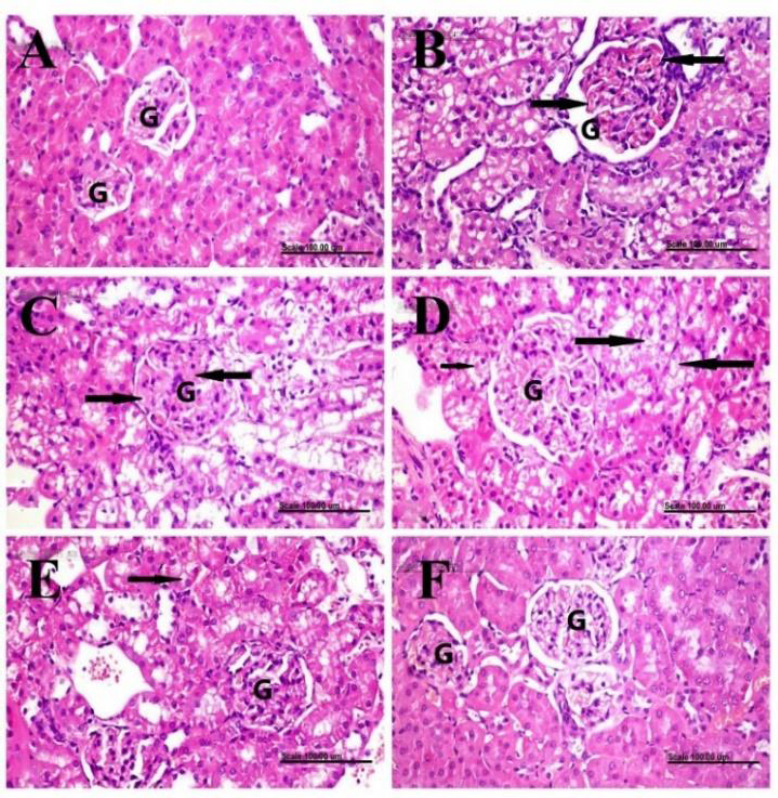
Fig. 4.

Histopathological examination and glomerular histomorphometry of renal tissue. Renal tissues of (A) normal control rats showing normal histology of renal glomeruli (G) and tubules; (B-D) doxorubicin-control rats showing glomerular congestion (arrows), mesangial matrix expansion (arrows), and pronounced swelling of the epithelial lining renal tubules with cytoplasmic vacuolization (long arrows) plus the presence of hyaline droplets (short arrow), respectively; (E) saxagliptin-treated group showing less swollen renal tubules with mild cytoplasmic vacuolization (arrow); and (F) vildagliptin-treated group showing a normal histological structure of glomeruli (G) and tubules. Stain, H&E, scale bar = 100 μm.
