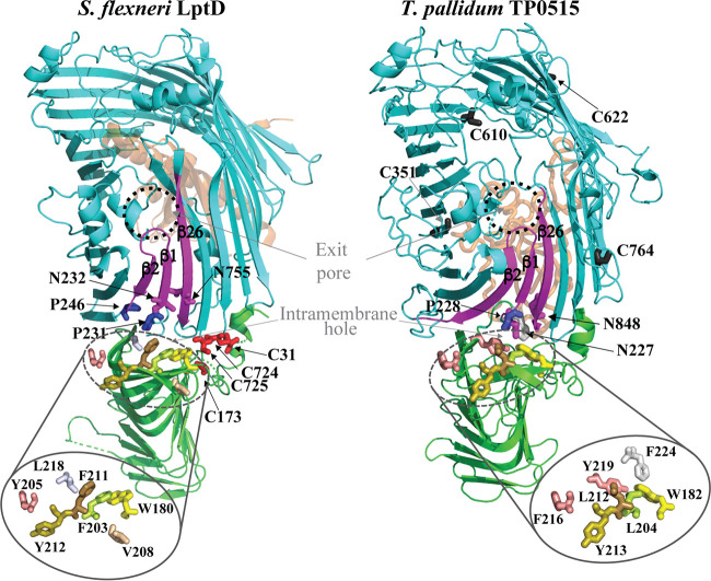
FIG 2.
Structural model of T. pallidum LptD (TP0515). Shown are cartoon diagrams of the LptD-LptE complex crystal structure (PDB accession number 4Q35) from Shigella flexneri (left) and the 3D structure model of the T. pallidum LptD ortholog (TP0515) (right). Both proteins are in the same orientation. The β-jelly roll N-terminal domain and 26-stranded β-barrel are shown in green and cyan, respectively. LptE of S. flexneri and the C-terminal extension of TP0515 are depicted as orange ribbons. With both proteins, the β-strands (β1 and β26) of the lateral gate and β2 are shown in magenta. Essential amino acids of the lateral gate (N232, P231, and P246) (38) and the intramembrane hole (W180, F203, Y205, V208, F211, and Y212) (75) and their T. pallidum TP0515 equivalents are displayed as sticks. Residues of intramembrane holes are zoomed-in for clarity. The four Cys residues required for the folding of S. flexneri LptD (76) are shown in red. Cys residues in T. pallidum LptD are shown as dark gray sticks.

