
FIGURE 2.
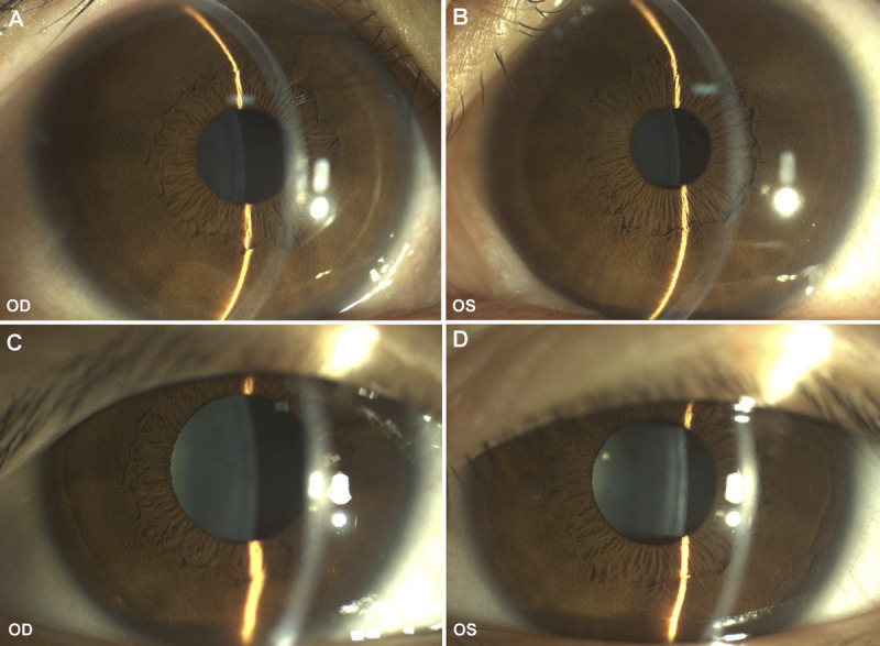
Anterior segment photographs of both eyes before and after treatment. (A) The anterior segment photograph of the right eye before treatment showed shallow peripheral anterior chamber. (B) The anterior segment photograph of the left eye before treatment showed shallow peripheral anterior chamber. (C) The peripheral anterior chamber of the right eye was deepened after treatment. (D) The peripheral anterior chamber of the left eye was deepened after treatment. OD = right eye; OS = left eye.