FIGURE 3.

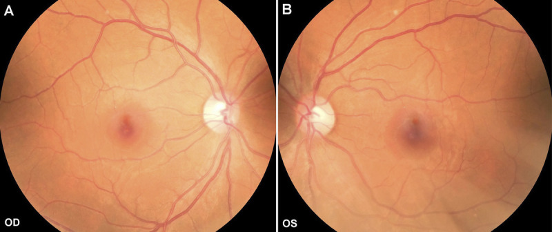
Fundus photographs of both eyes at first visit. (A) The fundus photograph of the right eye showed relatively normal fundus. (B) The fundus photograph of the left eye showed relatively normal fundus. OD = right eye; OS = left eye.

Fundus photographs of both eyes at first visit. (A) The fundus photograph of the right eye showed relatively normal fundus. (B) The fundus photograph of the left eye showed relatively normal fundus. OD = right eye; OS = left eye.