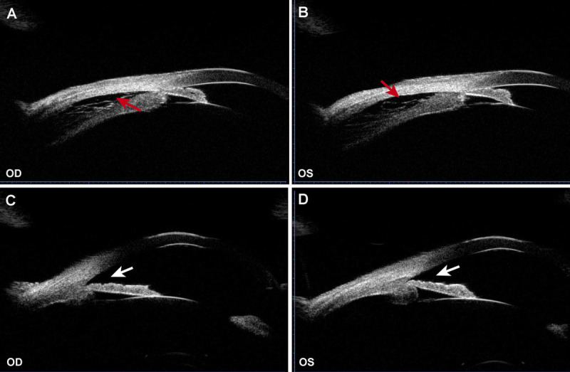
FIGURE 4.

The UBM examination of both eyes before and after treatment. (A) The UBM of the right eye before treatment revealed ciliary body detachment (red arrow) accompanied with angle closure (white arrow). (B) The UBM of the left eye before treatment revealed ciliary body detachment (red arrow) accompanied with angle closure (white arrow). (C) The ciliary body detachment of the right eye was resolved with angle opening (white arrow) after treatment. (D) The ciliary body detachment of the left eye was resolved with angle opening (white arrow) after treatment. OD = right eye; OS = left eye; UBM = ultrasound biomicroscopy.
