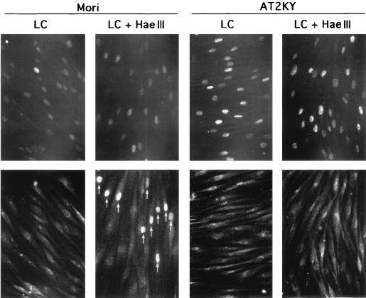
FIG. 7.
Phosphorylation at serine 15 of lactacystin-induced p53 protein following DNA double-strand breaks. Normal (Mori) and AT (AT2KY) fibroblasts were cultured for 3 h in the presence of lactacystin (50 μM) (LC), HaeIII (1 U/μl) was microinjected, and the cells were cultured for another 3 h in the presence of cycloheximide (20 μM) and lactacystin (50 μM) (LC + HaeIII). Fixed cells were double stained for p53 (top row) and phosphorylated serine 15 (bottom row); the same fields are shown. Perinuclear fluorescence was nonspecific staining due to the double staining. Arrows indicate microinjected cells containing p53 phosphorylated at serine 15. Note that lactacystin-induced p53 was phosphorylated at serine 15 following DNA double-strand breaks in normal cells, whereas p53 protein accumulated in AT cells was not phosphorylated after the same treatment.

