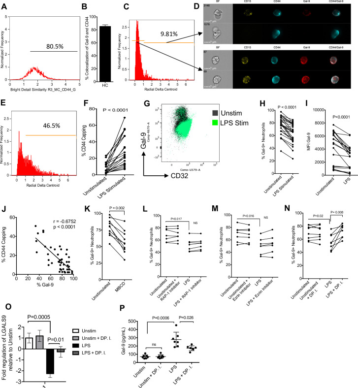
Fig 6. Gal-9 is shed by neutrophil mediated DP of CD44 upon activation.
(A) Percent colocalization of CD44 and Gal-9 on neutrophils quantified using an Amnis ImageStream. (B) Cumulative percent colocalization of Gal-9 and CD44 on the surface of neutrophils. (C) Representative plot of neutrophils with a high and low delta centroid XY. (D) Representative images of capped and dispersed CD44 on neutrophils. (E) Representative plot of LPS-treated neutrophil radial delta centroid. (F) Cumulative data of CD44 capping (delta centroid >1) in unstimulated and LPS-treated neutrophils. (G) Representative plot showing changing Gal-9 and CD32 expression on neutrophils untreated or stimulated with LPS. (H) Cumulative results showing the percent expression of surface Gal-9 on unstimulated and LPS stimulated neutrophils. (I) Cumulative results showing the MFI of surface Gal-9 on unstimulated and LPS stimulated neutrophils. (J) Cumulative data showing correlation between % Gal-9 expression on neutrophils and CD44 capping. (K) Surface expression of Gal-9 on neutrophils untreated or treated with MBCD. (L) Surface expression of Gal-9 on neutrophils untreated or treated with LPS in the presence or absence of a RAP-1 inhibitor, (M) ezrin inhibitor, and a (N) DP.I. (O) Cumulative data of Gal-9 mRNA expression and (P) shed Gal-9 in culture supernatants of LPS-activated neutrophils in neutrophils from HCs once stimulated with LPS for 3 hours in the presence or absence of DP.I. The underlying data can be found in S1 Data. DP, depalmitoylation; DP.I, depalmitoylation inhibitor; Gal-9, Galectin-9; HC, healthy control; LPS, lipopolysaccharide; MBCD, methyl-beta-cyclodextran; MFI, median fluorescence intensity.

