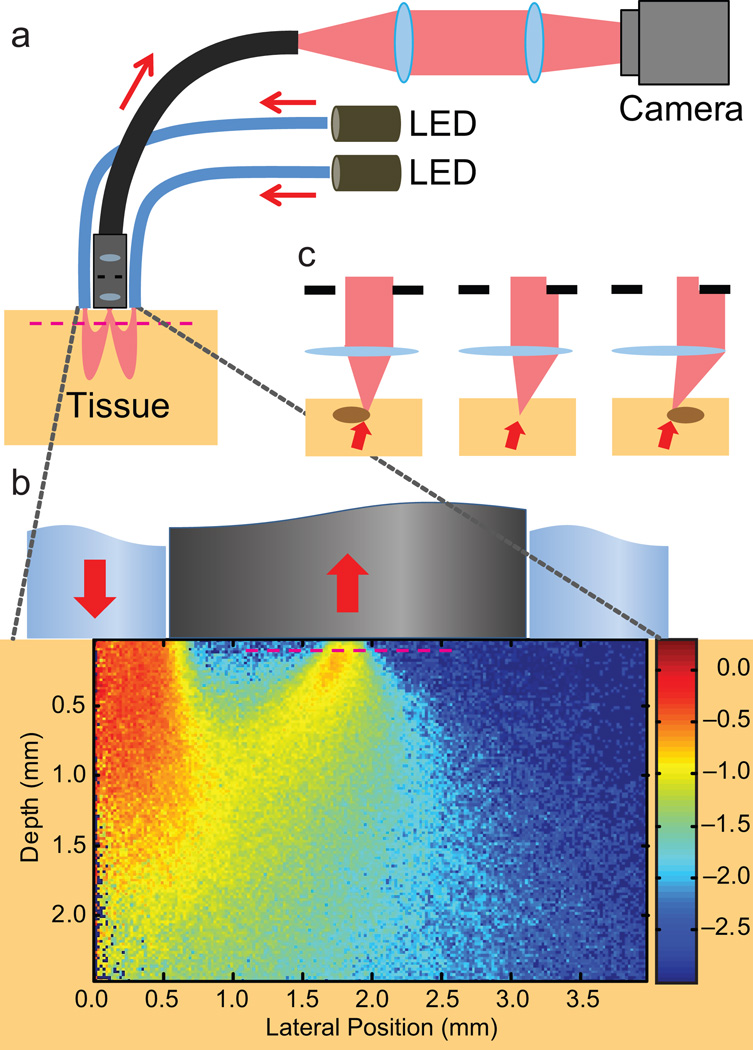
Figure 1.
An OBM setup with a contact-mode endomicroscope probe. (a) Illumination from two LEDs is sequentially delivered into a thick sample by diametrically opposed optical fibers attached to the probe housing. Multiple scattering in the sample redirects the light so that it trans-illuminates the focal plane of the probe micro-objective (magenta dashed line). An image from the focal plane is then relayed by a flexible fiber bundle and projected onto a digital camera. (b) Close-up of the probe distal end, onto which is superposed a density map obtained by Monte Carlo simulation of the light energy in the sample that was injected by a single fiber (from left) and collected by the micro-objective (Online Methods). Note the obliqueness of the light distribution through the focal plane. (c) Oblique trans-illumination leads to phase gradient contrast. With no phase gradient, oblique illumination is partially blocked by the aperture in the micro-objective (center panel). Phase gradients due to index of refraction variations at the focal plane tilt the light more or less (right/left) depending on the slope of the variations. As a result, more or less light is blocked by the micro-objective aperture, leading to intensity variations in the recorded image and hence phase gradient contrast4,5.

