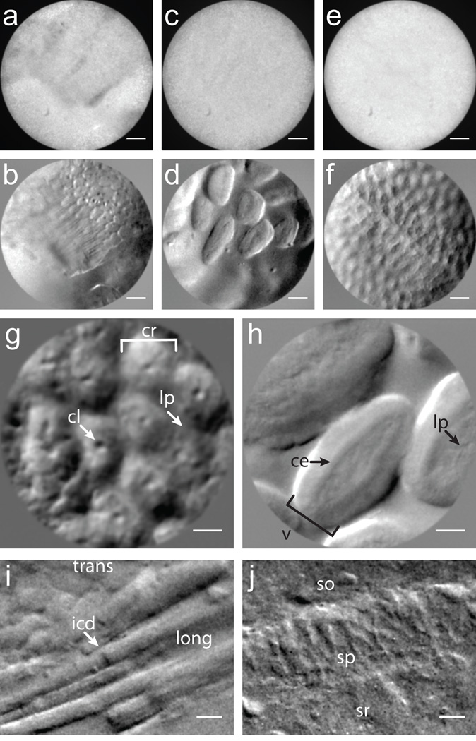
Figure 3.
Demonstration of OBM in excised mouse tissue. (a-f) Simultaneously acquired amplitude (a,c,e) and phase gradient (b,d,f) images of intestinal epithelium taken with a 1× micro-objective (field of view (FOV) 600 µm). (g,h) Higher magnification phase gradient images of the epithelium in the distal colon (g) and small intestine (h) were taken with a 2.5× micro-objective (FOV 240 µm). Crypts of Lieberkühn (cr), crypt lumens (cl) and lamina propia (lp) are indicated with arrows (g, Supplementary Video 6); ileal villi (v), columnar epithelium (ce) and lamina propia (lp) are indicated with arrows (h, Supplementary Video 7). (i) Transverse (trans) and longitudinal (long) aspects of mouse cardiac muscle tissue; intercalated discs (icd) are readily distinguished. (j, Supplementary Video 8) Mouse brain slice reveals pyramidal neurons in the CA1 region of the hippocampus. Stratum oriens (so), stratum pyramidale (sp) and stratum radiatum (sr) are apparent. Scale bars are (a-f) 75 µm, (g-h) 30 µm and (i-j) 20 µm.

