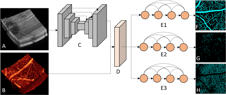
Fig. 1.
The architecture of the proposed method. (A) Structural OCT data volume. (B) OCTA data volume. (C) Three-dimensional convolutional module, with skip connections shown as arrows connecting the different layers. (D) The custom projection module. (E1-E3) The two-dimensional DenseNet convolutional modules. (F) Segmentation result for the superficial vascular plexus, (G) intermediate capillary plexus, and (H) deep capillary plexus.

