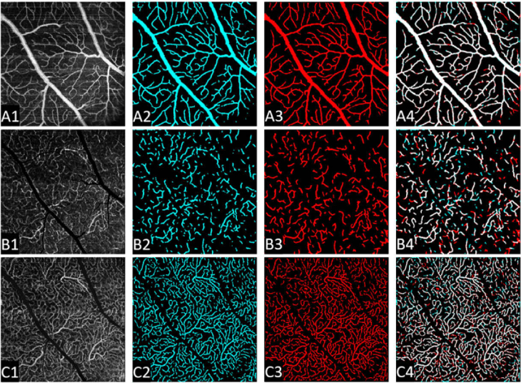
Fig. 6.
Vasculature segmentation results in all three retinal capillary plexuses. Row A: Superficial vascular plexus; Row B: intermediate capillary plexus; Row C: deep capillary plexus. First column: maximum projection en face angiograms. Second column: the vessel segmentation results from the proposed method. Third column: ground truth map. Last column: overlay of the ground truth and automated segmentation result. White indicates regions where the ground truth and automated segmentation results overlap.

