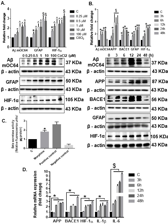
Figure 4.

Morphine-mediated amyloidosis and neuroinflammation in HPA. Western blot analysis showing dose-dependent upregulation of Aβ mOC64, GFAP, and HIF-1α (A) and time-dependent upregulation of Aβ mOC64, BACE1, GFAP, and HIF-1α (B) in HPAs exposed to morphine at the indicated dose and time points. (C) Morphine (500 nM)-exposed HPAs showed increased β-secretase activity by spectrofluorometric analysis. (D) qPCR showing increased expression of APP, BACE1, HIF-1α, IL1-β, and IL1-6 mRNAs in HPAs exposed to morphine (500 nM) at the indicated time points. GAPDH was used as an internal control for mRNA expression. Data are presented as mean±SEM. One-way ANOVA followed by Bonferroni post hoc test was performed, $P< 0.001, ^P< 0.01, *P < 0.05 versus control. Abbreviations: APP, amyloid precursor protein, GFAP, glial fibrillary acidic protein, HIF-1α, hypoxia-inducible factor 1α, 1IL, interleukin, qPCR, quantitative polymerase chain reaction.
