FIGURE 2.

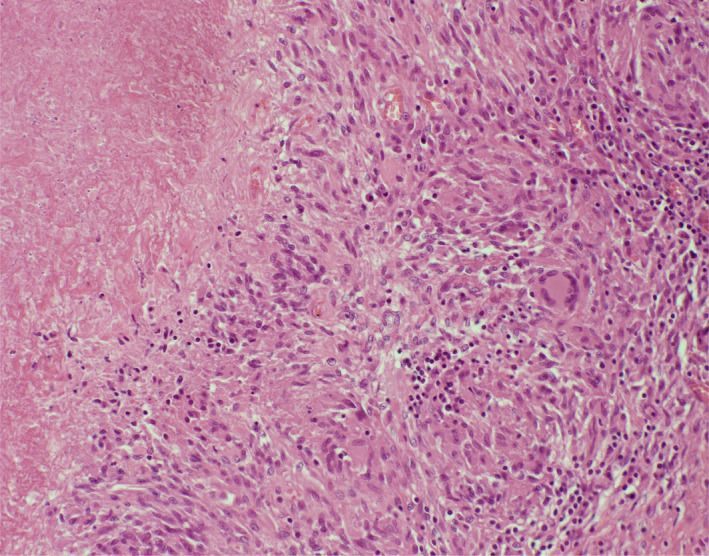
Microscopic examination of the perianal fistula showing caseous necrosis surrounded by epithelioid cells and giant cells

Microscopic examination of the perianal fistula showing caseous necrosis surrounded by epithelioid cells and giant cells