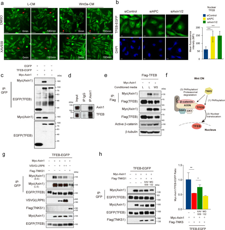
Fig. 2. Wnt signaling dependent nuclear localization of TFEB is regulated in the β-catenin destruction complex.
a Treatment of XAV939 inhibits Wnt3a-mediated nuclear localization of TFEB. TFEB-EGFP stable cells were treated with or without 2 μM XAV939 in L-CM or Wnt3a-CM. Scale bar, 50 μm. b Knockdown of the destruction complex component induced nuclear localization of TFEB-EGFP. Immunofluorescence analysis was performed in TFEB-EGFP stably expressing cells transfected with control, Axin1/2 or APC siRNA. Quantification of mean fluorescence intensity (MFI) of TFEB in the nuclear ROI region from 8 bit confocal images (maximum gray value, 256) is shown in right panel. Scale bar, 20 μm. c TFEB interacts with Axin1 which is a component of the β-catenin destruction complex. EGFP-TFEB and Myc-Axin1 were co transfected into HEK293T cells. Cell lysates were then immunoprecipitated with anti-GFP antibody and immunoblotted with the indicated antibodies. d Axin interacts with TFEB at the endogenous level. HeLa cell lysates were immunoprecipitated with endogenous anti-Axin1 antibody and immunoblotted with endogenous anti-TFEB antibody. e, g, and h Activation of Wnt signaling reduced interaction between TFEB and Axin1. e Flag-TFEB and Myc-Axin1-expressing HEK293T cells were treated with Wnt3a-CM, followed by immunoprecipitation with anti-Flag antibody and immunoblotting with the indicated antibodies. f Schematic diagram for the prediction of effects of TNKS1 overexpression on dissociation of TFEB from the β-catenin destruction complex. g TFEB-EGFP and Myc-Axin1-expressing HEK293T cells were co-transfected with VSVG-LRP6 or Flag-TNKS1. Lysates were immunoprecipitated with anti-GFP antibody and immunoblotted with the indicated antibodies. h Treatment of XAV939, but not MG132, blocked TNKS1 dependent reduction of interaction between TFEB and Axin1. TFEB-EGFP and Myc-Axin1 and Flag- TNKS1 expressing HEK293T cells were treated with XAV939 or MG132. Cell lysates were immunoprecipitated with anti-GFP antibody and immunoblotted with the indicated antibodies. Quantification of the ratio of immunoprecipitated Myc-Axin1/TFEB-EGFP of three independent immunoblots was shown in the right panel. Data information: In (b) and (h), data are presented as mean ± SEM. *P < 0.05, **P < 0.01, and ***P < 0.005 (Student’s t test).

