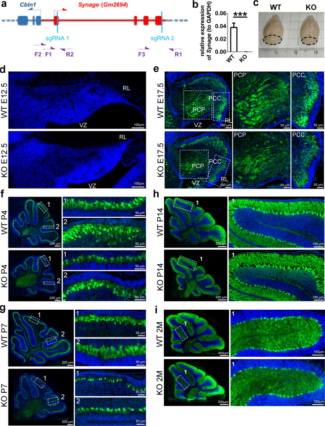
Fig. 2. Synage knockout mice show cerebellar defects and neuronal loss during cerebellar development.
a Schematic representation of the position of Synage, Cbln1, sgRNAs, and genotyping primers (F forward primers, R reverse primers). b Relative expression levels of Synage in the cerebella of WT and knockout (KO) adult mice detected by RT-qPCR. c Gross morphology of representative brains from adult WT and KO male mice. d Hoechst 33342 staining of cerebella from E12.5 WT and KO mice (WT, n = 9; KO, n = 6). e–i Immunofluorescence staining of the Purkinje cell marker protein (Calbindin, green) in the cerebella from E17.5 (e), P4 (f), P7 (g), P14 (h), and 2-month-old (i) WT and KO mice. WT (E17.5), n = 7; KO (E17.5), n = 3; WT (P4, P7, P14, and 2 months), n = 3; KO (P4, P7, P14, and 2 months), n = 3. Nuclei were stained with Hoechst 33342 (blue).

