FIGURE 4.

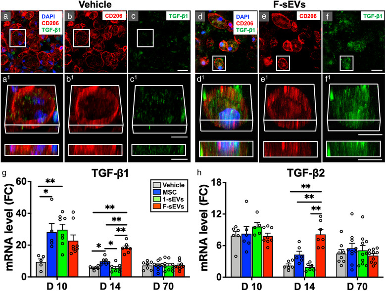
Both infusion of MSCs and fractionated dosing of MSC‐sEVs upregulate TGF‐β in the injured spinal cord. a‐f, Confocal micrographs of representative regions of frozen sectioned contused spinal cord harvested 14‐day post‐SCI and 7 days after the treatment with Vehicle (a‐c, left) or fractioned MSC‐sEVs (d‐f, right), immunostained with antibodies directed against Type M2 macrophage marker CD206 (red), and TGF‐β1 (green), and counterstained with DAPI (blue). Images in each group (a‐c, and d‐f) from left to right show the same area with fluorescence channels for CD206, TGF‐β1, & DAPI (a and f), CD206 (b and e), and TGF‐β1 (c and f). Note that TGF‐β1 staining is strongly expressed and localized within macrophages staining positive for the M2 type marker in the F‐sEVs group, while such staining is negligible in the Vehicle group. Scale bars indicate 20 μm (c and f) and 10 μm (c1 and f1). g and h, Graphs illustrating the qRT‐PCR analysis of the relative expression of mRNAs for TGF‐β1 (g) and TGF‐β2 (h). [The ΔCT was calculated against the internal control (GAPDH), and ΔΔCT (mRNA level) was calculated against the ΔCT of the control.] Note the transient upregulation of TGF‐β1 at 10‐day post‐ SCI in the 1‐sEVs treatment group, compared with the upregulation of TGF‐β1 and TGF‐β2 expression in both the MSC and the F‐sEVs treatment groups at 14‐day post‐SCI. Values are presented as means ± SEM. A 1‐way ANOVA followed by the Tukey‐Kramer test or the Kruskal‐Wallis test followed by the Steel‐Dwass test was conducted. *: P < .05, **: P < .01, TGF: transforming growth factor, MSC: mesenchymal stem/stromal cell, MSC‐sEVs: small extracellular vesicles derived from MSCs, TGF‐β: transforming growth factor‐beta, DAPI: 4′,6‐diamidino‐2‐phenylindole, qRT‐PCR: quantitative reverse transcription‐polymerase chain reaction, ΔCT: delta‐cycle threshold, SCI: spinal cord injury, FC: fold change
