
Figure 5 .
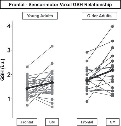
Higher GSH levels in sensorimotor versus frontal cortex. Frontal and SM (sensorimotor) GSH levels within each young (left) and older (right) adult. Each line represents one participant. For both age groups, GSH levels (group medians shown in black) were higher in the sensorimotor compared with the frontal voxel.