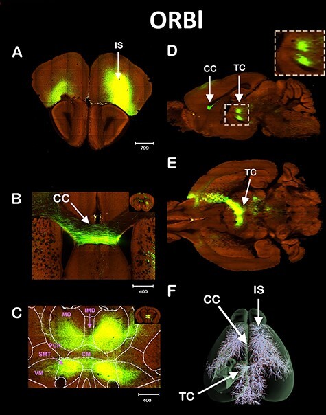
Figure 2 .

ORBl Interhemispheric thalamic connectivity. Tract tracing of the ORBl cortex. IS shown in (A), Coronal view of the CC crossing (B), and of the TC (C). Sagittal (D) and axial (E) view of the TC, and the 3D reconstruction (F). The following thalamic nuclei were labeled only in the contralateral hemisphere in order to not obstruct the image. VM = ventromedial and SMT = submedial. Scale bars in μm.
