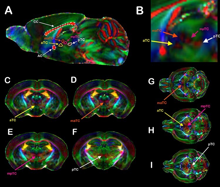
Figure 3 .

TC revealed by DWI. (A) Direction color encoded (DEC) sagittal DWI maps of a C57bl6/J mouse brain showing the CC, AC, and the TC. In all animals, we were able to distinguish at least 4 distinct TCs, named according to their relative disposition along the anteroposterior axis: the anterior TC (aTC, yellow arrows), the medial anterior TC (maTC, orange arrows), the medial posterior TC (mpTC, magenta arrows), and the posterior TC (pTC, white arrows). (B) Inset showing the TCs in greater detail. Coronal (D–F) and axial (G–I) DWI planes showing the TCs crossing the midline. In the DEC DWI maps, red represents mediolateral (ML) diffusion, green represents anteroposterior (AP) diffusion, and blue represents dorsoventral (DV) diffusion.
