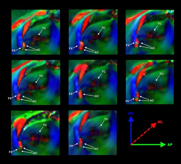
Figure 5 .

Commissural abnormalities in Balb/c. Schematic anatomical drawing of a mouse brain midsection highlighting the interhemispheric commissures, CC, AC, TC, and the fornix (FX) (A). Direction color encoded (DEC) sagittal DWI map of 8 Balb/C mouse brains showing the anatomical variability of the AC, fornix (FX), and TC. Interestingly, in most (7/8) Balb/c mice, the FX transects the AC (B).
