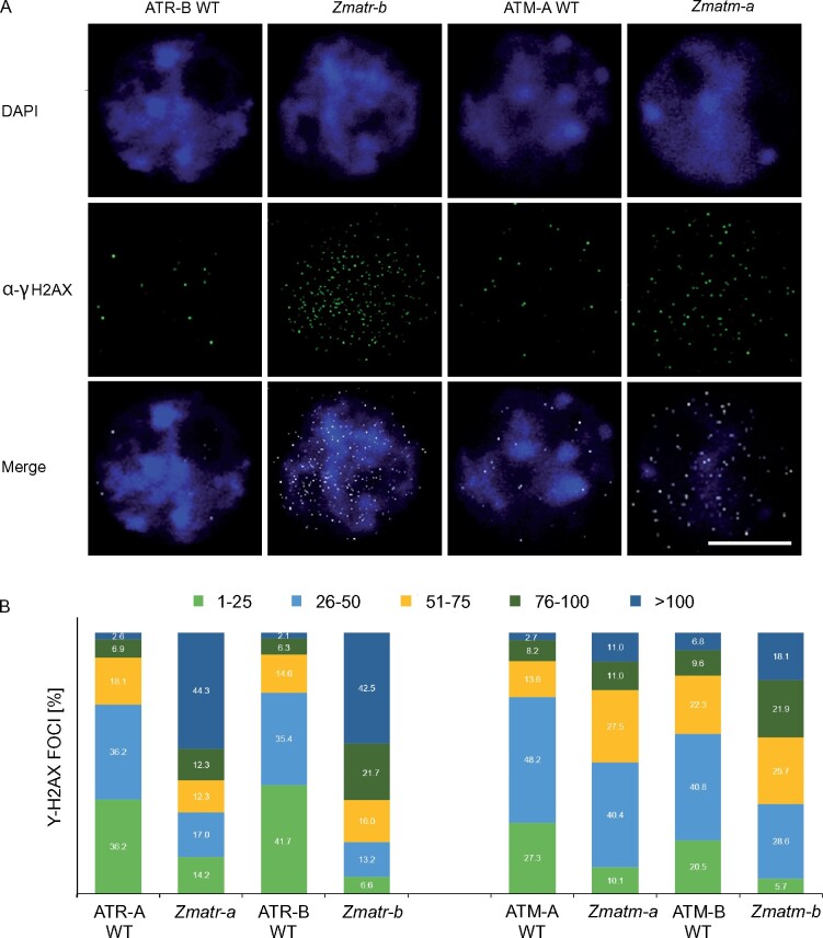
Figure 7.
Detection of γH2AX foci in Zmatr and Zmatm mutant embryos. A, Immunostaining of γH2AX foci accumulation (green) in nuclei stained with DAPI (blue) of ATR WT, Zmatr, ATM-WT, and Zmatm embryos at 16 DAP. A representative nucleus is shown for each line. Scale bar = 5 µm. B, Quantification of γH2AX foci in ATR-WT, Zmatr, ATM-WT, and Zmatm embryos at 16 DAP. For each sample, the γH2AX foci of 100 nuclei were counted and grouped into five categories: 1–25, 26–50, 51–75, 76–100, and >100 foci per nucleus. Two independent lines were analyzed.

