Figure 4 .

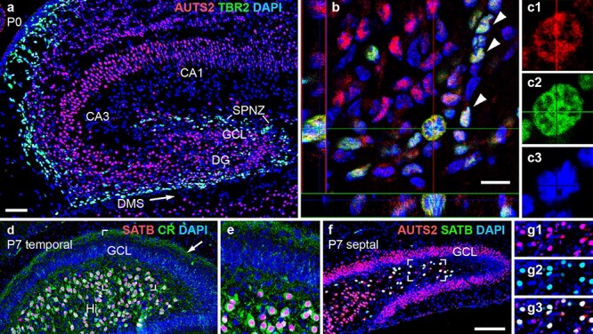
AUTS2 expression in neonatal hippocampus. (a–c3) Double IF for AUTS2 (red) and TBR2 (green) on P0. In (b), a confocal image of the DMS with orthogonal views (left and bottom) shows AUTS2 expression in TBR2+ INPs (arrowheads), including an M-phase INP (crosshairs) shown at higher magnification in (c1–c3). (d–e) Double IF for SATB1/2 (red) and calretinin (green) in P7 DG at temporal level. Calretinin+ recurrent axon collaterals from HMNs are indicated in the DG inner molecular layer (arrow). The boxed area in (d) is shown at higher magnification in (e). (f–g3) Double IF for SATB1/2 (green) and AUTS2 (red) in P7 DG at septal level. The boxed area of the Hi in (f) is shown at higher magnification in (g1–g3). Sagittal sections with DAPI (blue) counterstain. Scale bars: a, 50 μm for a, d, and 25 μm for e; b, 10 μm for b, and 5 μm for c1–c3; f, 100 μm for f, and 50 μm for g1–g3.
