
Figure 6 .
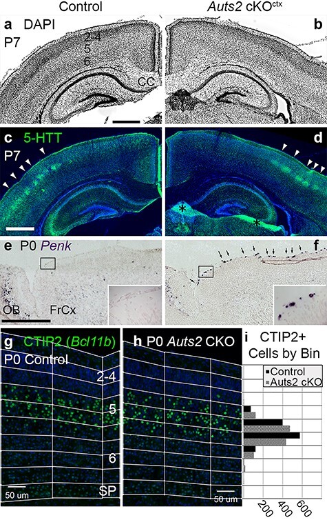
Normal structure but molecular defects in Auts2 cKOctx neocortex. (a, b) DAPI staining (inverted fluorescence) of P7 control (a) and Auts2 cKOctx (b) brains. Neocortical layers are numbered in (a). Coronal sections. Abbreviation: CC, corpus callosum. (c, d) IF to detect serotonin transporter (5-HTT) in same sections as (a, b). Arrowheads indicate 5-HTT+ layer 4 somatosensory barrels. Asterisks in (d): tissue artifacts. (e, f) ISH to detect Penk in P0 control (e) and Auts2 cKOctx (f) brains. Boxed areas are shown at higher magnification in insets. Arrowheads in (f) indicate cells with increased Penk expression, presumably C-R neurons. Sagittal sections. Abbreviations: FrCx, frontal cortex; OB, olfactory bulb. (g, h) IF to detect CTIP2 (gene Bcl11b) in P0.5 control (g) and Auts2 cKOctx (h) neocortex. Neocortical layers are numbered in (g). Pial to subplate binning is as indicated. Sagittal sections through somatosensory cortex. (i) Ctip2+ cells quantified across 9 binned images each for control and Auts2 cKO (wilcox-test P = 0.5321). Abbreviations: IZ, intermediate zone; SP, subplate. Scale bars: a, 500 μm for a–d; e, 500 μm for e, f; and g, 100 μm for g, h.