
FIGURE 1.
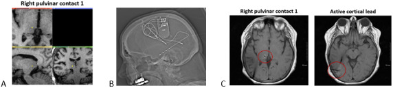
Postoperative Images from Patient 3. (A) Depth lead placement in the pulvinar (B) Post‐implantation CT scan (C) Axial views of right pulvinar contact 1 and right cortical depth lead in the occipitotemporal lobe
Official websites use .gov
A
.gov website belongs to an official
government organization in the United States.
Secure .gov websites use HTTPS
A lock (
) or https:// means you've safely
connected to the .gov website. Share sensitive
information only on official, secure websites.
Postoperative Images from Patient 3. (A) Depth lead placement in the pulvinar (B) Post‐implantation CT scan (C) Axial views of right pulvinar contact 1 and right cortical depth lead in the occipitotemporal lobe