
FIGURE 2.
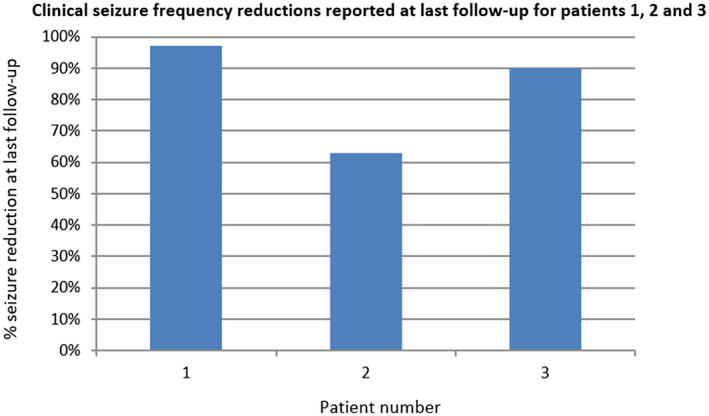
Clinical seizure frequency reductions reported at last follow‐up. Patient 2 had a right pulvinar depth and a right parietal depth, patient 3 a right pulvinar depth and right occipitotemporal depth, and patient 1 had two RNS Systems placed, with bilateral pulvinar depth leads and a right occipital cortical strip and a left occipital depth lead