Figure 1.

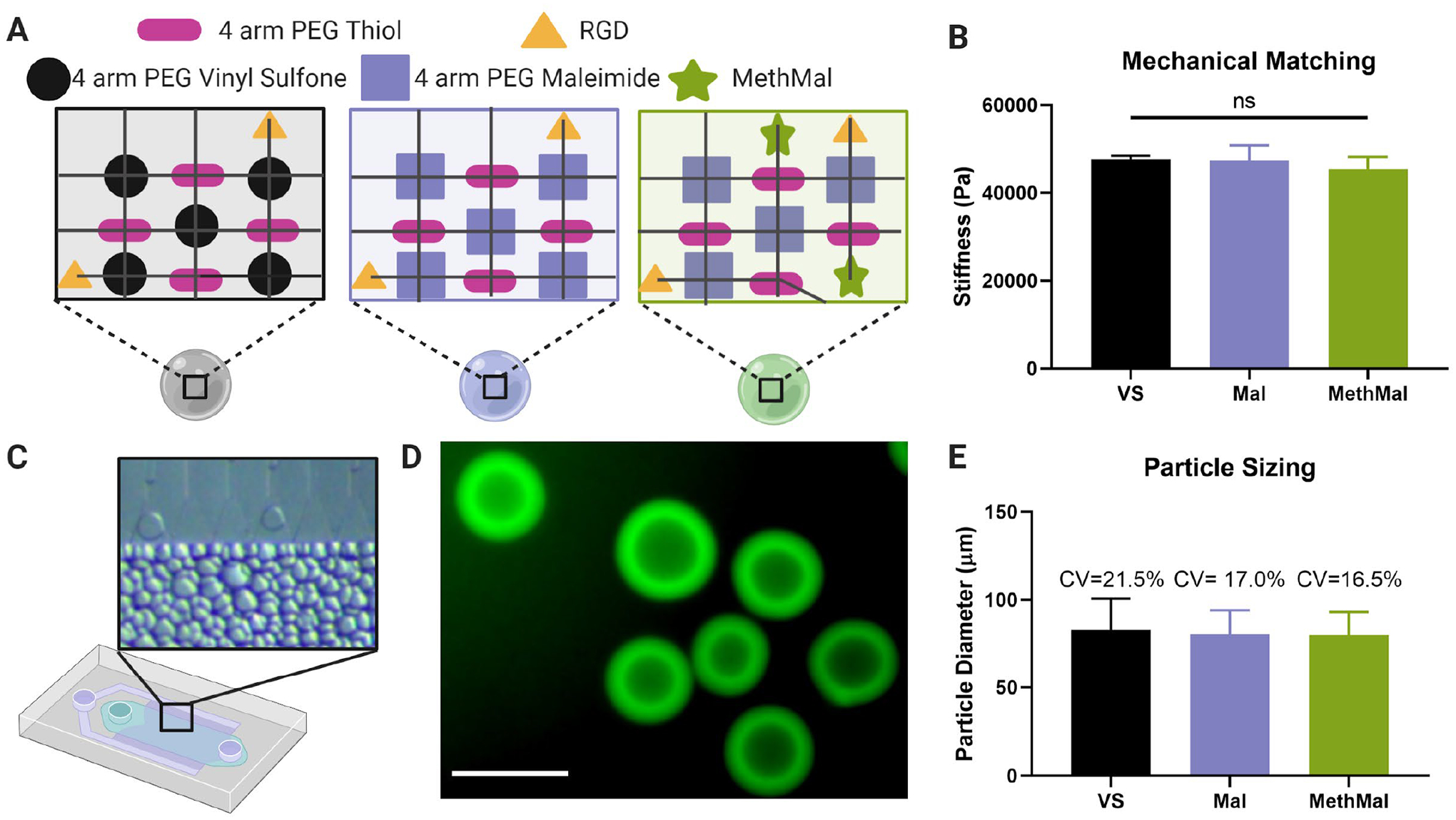
Synthesis and characterization of three microgel types. A) The three gel formulations were composed of a PEG-Vinyl Sulfone backbone (VS), a PEG-Maleimide backbone (Mal), and a PEG-Maleimide with 1mM MethMal (MethMal). All gel formulations were crosslinked with a 4-arm PEG Thiol and had RGD cell adhesive peptide. B) The three gel formulations were mechanically matched to have a Young’s modulus of ~46kPa determined by Instron testing of macroscale gels. Note: All microgel compositions were formulated to stoichiometrically provide a theoretical 1mM excess photoannealing functional group for all conditions. C) Microgels were produced using a high throughput microfluidics device. D) Biotinylated microgels were fluorescently visualized with streptavidin-488. Scale bar represents 100μm. E) Microgels were size matched to approximately ~75μm in diameter. All graphs show mean +/− standard deviation. One-way ANOVA was used to determine significance in mechanical moduli.
