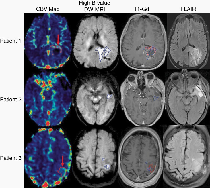
Fig. 1.
In the top row, representative images from Patient 1 demonstrate hyperperfused tumor (red arrow, first column) identified with dynamic contrast-enhanced MRI, and hypercellular tumor (blue volume, second column) identified with high b value diffusion-weighted MRI. Both volumes are overlayed on the T1-weighted gadolinium-enhanced MRI (third column), and the non-enhancing hypercellular tumor extends across the splenium. In the middle row, the left temporal lobe tumor from Patient 2 demonstrates residual, non-enhancing hypercellular tumor posterior to the surgical cavity (blue volume, second column) without corresponding hyperperfusion. This non-enhancing tumor region is significantly smaller than the nonspecific FLAIR hyperintensity surrounding the cavity (last column). In the bottom row, representative images from Patient 3 demonstrate both hyperperfused (red arrow, first column) and hypercellular (blue volume, second column) tumor in the left posterior frontal lobe that was non-enhancing and superior to the surgical cavity. Abbreviations: CBV, cerebral blood volume; DW-MRI, diffusion-weighted magnetic resonance image; FLAIR, fluid-attenuated inversion recovery; T1-Gd, gadolinium-enhanced T1-weighted MRI.

