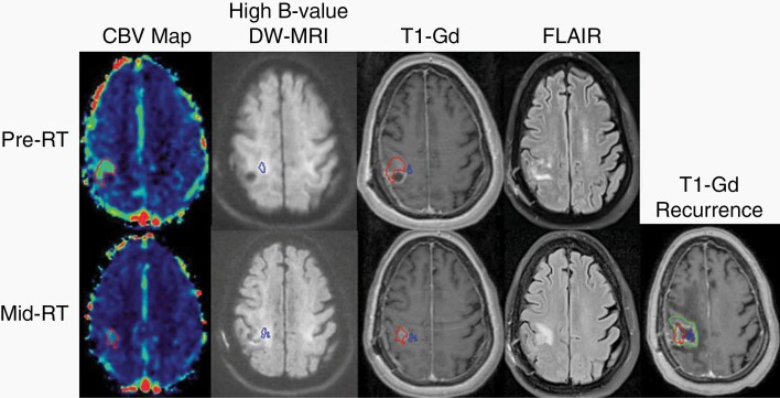
Fig. 3.
Correlation between persistent and developing mid-radiation hyperperfused and hypercellular tumor volumes and tumor recurrence. A hyperperfused region of tumor (red volume, far left column) extending outside of the enhancing target (third column, upper panel) demonstrates resolution laterally but persistence medially by mid-RT. A geographically distinct but also non-enhancing hypercellular region of tumor (blue volume, second column) demonstrates persistent but also newly developing regions of tumor further medially by mid-RT, while the enhancing target regresses (third column, bottom panel). Both persistent and developing hyperperfused and hypercellular tumor volumes detected mid-radiation correspond to the eventual tumor recurrence (green volume, far right panel) 8 months after chemoradiation. Abbreviations: CBV, cerebral blood volume; DW-MRI, diffusion-weighted magnetic resonance image; FLAIR, fluid-attenuated inversion recovery; RT, radiation; T1-Gd, gadolinium-enhanced T1-weighted MRI.

