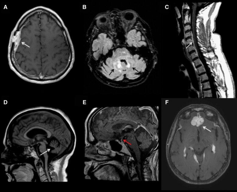
Fig. 2.
Magnetic resonance imaging (MRI) of histiocytoses of the nervous system. (A) Axial T1 post-gadolinium MRI demonstrates calvarial-dural Langerhans cell histiocytosis (LCH). (B) Axial T2 FLAIR (fluid-attenuated inversion recovery) MRI demonstrates Erdheim-Chester disease (ECD) of the brainstem and cerebellum. (C) Sagittal T1 post-gadolinium mixed histiocytosis (ECD with Rosai-Dorfman-Destombes disease [RDD]) of the spine. (D) Sagittal T1 MRI demonstrates profound cerebellar atrophy in neurodegenerative LCH. (E) Sagittal T1 post-gadolinium MRI with enhancement and thickening of the infundibulum in a patient with LCH. (F) Axial T1 post-gadolinium MRI with RDD involving dura and bifrontal lobes.

