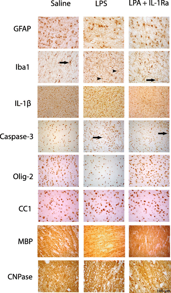
Fig. 6.

Representative photomicrographs showing positive staining of GFAP, Iba-1, IL-1β, caspase 3, Olig-2, CC1, MBP and CNPase in the periventricular white matter tracts. Scale bar = 100 μm. Arrows in the Iba-1 photomicrographs indicate microglia displaying a resting ramified phenotype, characterised by a small cell body with > 1 branching process. Arrowheads indicate microglia displaying an amoeboid morphology, characterised by a large cell body with ≤ 1 branching process. Arrows in the caspase-3 photomicrographs indicate positive cells displaying both staining and apoptotic bodies
