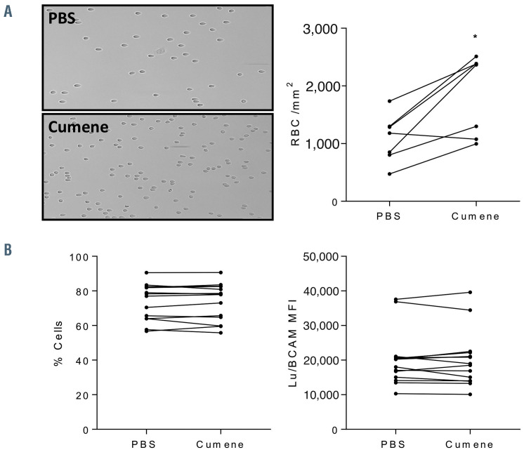
Figure 3.
Effect of in vitro oxidation on AA red blood cell adhesion to laminin. (A) Left panel: typical microscopy images of non-oxidized and cumene hydroperoxideoxidized AA red blood cells (RBC) adhering to Laminin 521 at 3 dyn/cm2. Right panel: quantification of cell adhesion showing the mean number of adherent RBC/mm2 in seven oxidized and non-oxidized AA RBC samples at 3 dyn/cm2. Wilcoxon test, *P<0.05. (B) Flow cytometry analysis of Lu/BCAM expression at the RBC surface expressed as percentage of Lu/BCAM-positive RBC (left panel) and mean fluorescence intensity (MFI) of these RBC (right panel) under non-oxidized (phosphate buffered saline [PBS]) and oxidized conditions (cumene). No significant differences were observed, Wilcoxon test, P=0.0714.

