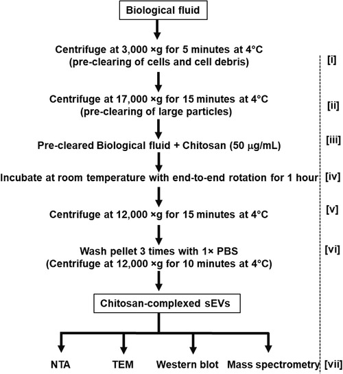
FIGURE 1.

Flow chart for chitosan‐based small extracellular vesicle (sEV) isolation. Biofluids are collected and precleared in two steps; first by centrifugation at 3,000 × g for 5 min at 4°C [i], and a second centrifugation at 17,000 × g for 15 min at 4°C [ii]. Chitosan is added to a final concentration of 50 μg/ml [iii] and samples are incubated at room temperature for 1 h with end‐to‐end rotation to form chitosan‐sEV complexes [iv]. The chitosan‐sEV complexes are pelleted by centrifugation at 12,000 × g for 15 min at 4°C [v]. The resulting pellet of chitosan‐sEV complex is washed 3 times with 1 × PBS by centrifugation at 12,000 × g for 10 min at 4°C [vi]. Chitosan‐sEV complexes are used for various downstream analyses [vii], such as nanoparticle tracking analysis (NTA), transmission electron microscopy (TEM), Western blot and mass spectrometry
