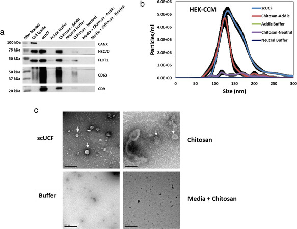
FIGURE 2.

Chitosan‐mediated sEV isolation from HEK‐293 conditioned culture media (CCM). (a) Western blot analyses of the canonical EV markers CD63, CD9, HSC70, and FLOT1 for material isolated by the addition of acidic or neutral formulations of 60–120 kDa chitosan at a final concentration of 50 μg/ml to 2 ml of CCM. sEVs isolated using scUCF from 2 ml of CCM were used as a positive control and the acidic or neutral buffers in which chitosan was dissolved were included as negative controls. Non‐conditioned media (Media) was also used to evaluate whether any sEV artefacts are present within the chitosan preparation. Blots were also probed for CANX, which is absent from sEVs; total cell lysate from MCF‐10A cells was used as a positive control for CANX. (b) NTA analysis of sEVs isolated from 2 ml of HEK‐293 CCM by scUCF, the acidic formulation of chitosan, the neutral formulation of chitosan, and buffer controls. Particle concentration (particles/ml) is plotted against size (nm) of particles. The mean and the standard error of the mean (n = 3) are shown. (c) TEM images of sEVs isolated by scUCF, chitosan (acidic formulation), and buffer from HEK‐293 CCM. Non‐conditioned media incubated with the acidic formulation of chitosan was also used to confirm that chitosan does not aggregate into sEV‐sized particles. Scale bars represent 200 nm. Membrane‐bound structures consistent with sEVs are indicated with white arrows
