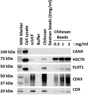
FIGURE 7.

sEV isolation by chitosan‐coated magnetic beads. Increasing amounts of chitosan‐coated magnetic beads (0.5, 1, and 2 mg/ml) were added to 1 ml of HEK‐293 CCM; dextran‐coated magnetic beads (2 mg/ml) or buffer served as a negative controls and scUCF or the addition of the acidic formulation of chitosan to a final concentration of 50 μg/ml were used as positive controls. Western blot analyses of canonical EV markers CD63, CD9, HSC70, and FLOT1, as well as the non‐sEV marker CANX were performed for material isolated from HEK‐293 CCM; total cell lysate from HEK‐293 cells was used as a positive control for CANX
