. 2021 Jul 6;300(3):681–689. doi: 10.1148/radiol.2021204441

Figure 4:

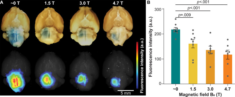
Evans blue delivery via focused ultrasound combined with microbubble-induced blood-brain barrier opening in different magnetic fields. (A) Representative photographs (top row) and corresponding fluorescence images (bottom row) of mouse brains treated in magnetic fields of approximately 0 T, 1.5 T, 3.0 T, and 4.7 T. (B) Fluorescence intensity quantification of mice treated in magnetic fields of approximately 0 T, 1.5 T, 3.0 T, and 4.7 T. Error bars indicate standard error of the mean.

Evans blue delivery via focused ultrasound combined with microbubble-induced blood-brain barrier opening in different magnetic fields. (A) Representative photographs (top row) and corresponding fluorescence images (bottom row) of mouse brains treated in magnetic fields of approximately 0 T, 1.5 T, 3.0 T, and 4.7 T. (B) Fluorescence intensity quantification of mice treated in magnetic fields of approximately 0 T, 1.5 T, 3.0 T, and 4.7 T. Error bars indicate standard error of the mean.