. 2021 Jul 6;300(3):681–689. doi: 10.1148/radiol.2021204441

Figure 5:

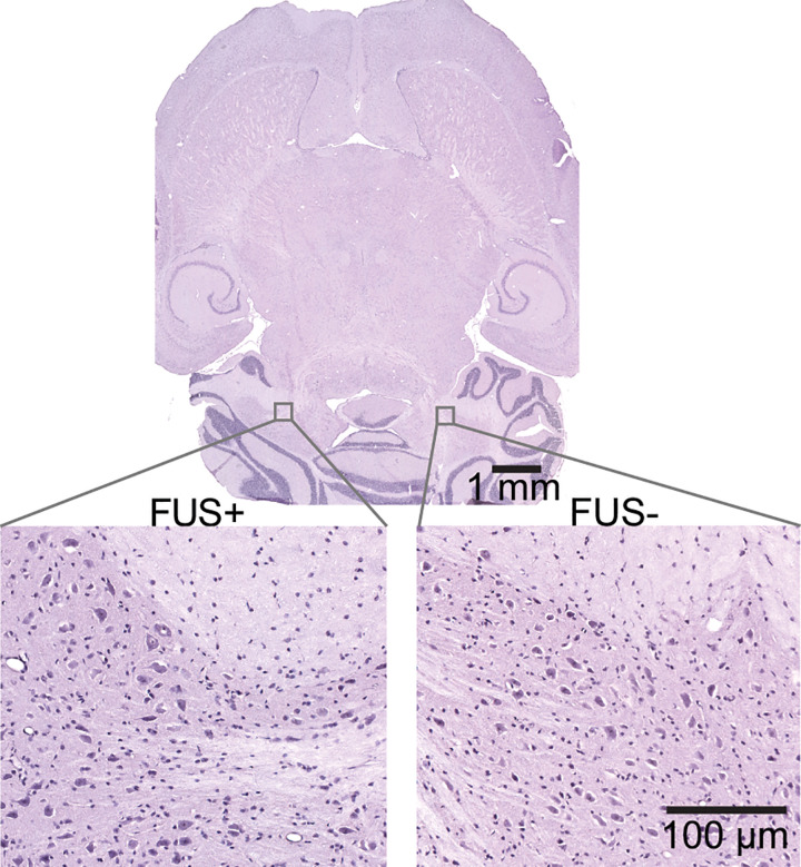
Hemotoxylin-eosin–stained horizontal whole-brain slice (top panel) with higher-magnification images (bottom panels) obtained from the focused ultrasound–treated brain region (FUS+) and contralateral untreated control region (FUS−). No gross tissue damage was observed on the whole-brain slice. Higher-magnification images showed that erythrocyte extravasation and neuronal damage were not observed in either the focused ultrasound–treated brain region or the untreated control brain region.

Hemotoxylin-eosin–stained horizontal whole-brain slice (top panel) with higher-magnification images (bottom panels) obtained from the focused ultrasound–treated brain region (FUS+) and contralateral untreated control region (FUS−). No gross tissue damage was observed on the whole-brain slice. Higher-magnification images showed that erythrocyte extravasation and neuronal damage were not observed in either the focused ultrasound–treated brain region or the untreated control brain region.