. 2021 Jun 22;300(3):626–632. doi: 10.1148/radiol.2021204500

Figure 1:

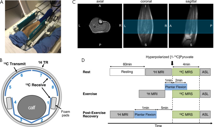
Experimental set-up and study protocol. A, Photograph depicts positioning of calf muscle in carbon 13 (13C)–hydrogen 1 (1H) dual-frequency radiofrequency coil before connecting anterior part of coil. B, Illustration shows how calf muscle was wrapped with flexible posterior 13C array receiver coils (channels 1–4). TR = transmit and receive. C, Localized single-shot fast spin-echo 1H MRI scans obtained using radiofrequency coil. Blue region indicates prescribed axial slab for 13C MR spectroscopy (10-cm thick). A = anterior, I = inferior, L = left, P = posterior, R = right, S = superior. D, Diagram shows how dynamic 13C MR spectroscopy (MRS) was performed at three metabolic states—rest, exercise, and recovery. ASL = arterial spin labeling.

Experimental set-up and study protocol. A, Photograph depicts positioning of calf muscle in carbon 13 (13C)–hydrogen 1 (1H) dual-frequency radiofrequency coil before connecting anterior part of coil. B, Illustration shows how calf muscle was wrapped with flexible posterior 13C array receiver coils (channels 1–4). TR = transmit and receive. C, Localized single-shot fast spin-echo 1H MRI scans obtained using radiofrequency coil. Blue region indicates prescribed axial slab for 13C MR spectroscopy (10-cm thick). A = anterior, I = inferior, L = left, P = posterior, R = right, S = superior. D, Diagram shows how dynamic 13C MR spectroscopy (MRS) was performed at three metabolic states—rest, exercise, and recovery. ASL = arterial spin labeling.