Figure 1.

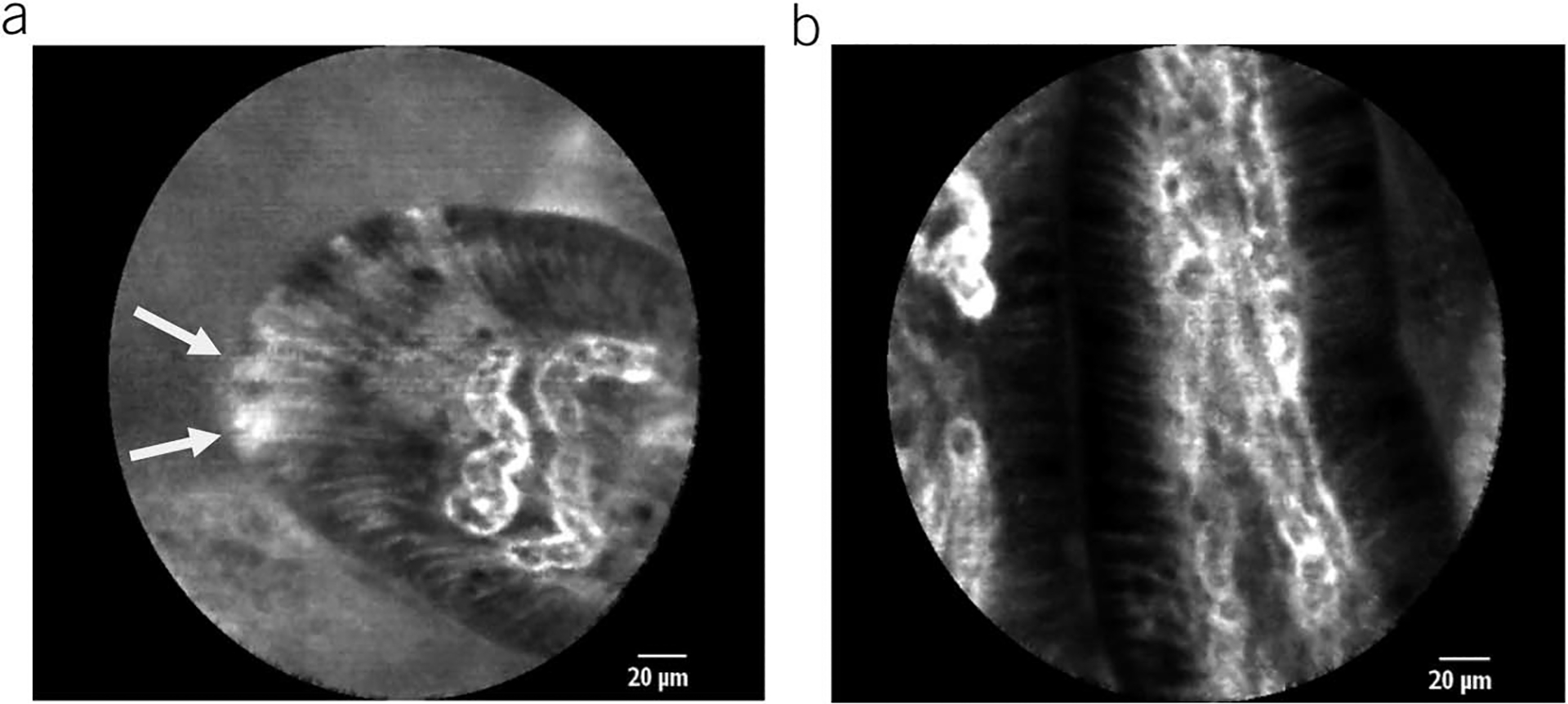
Representative probe-based CLE images of duodenal villi: (a) a patient with functional dyspepsia with several adjacent epithelial gaps (white arrowheads indicating epithelial gaps); (b) healthy individual without epithelial gaps.

Representative probe-based CLE images of duodenal villi: (a) a patient with functional dyspepsia with several adjacent epithelial gaps (white arrowheads indicating epithelial gaps); (b) healthy individual without epithelial gaps.