Figure 7.

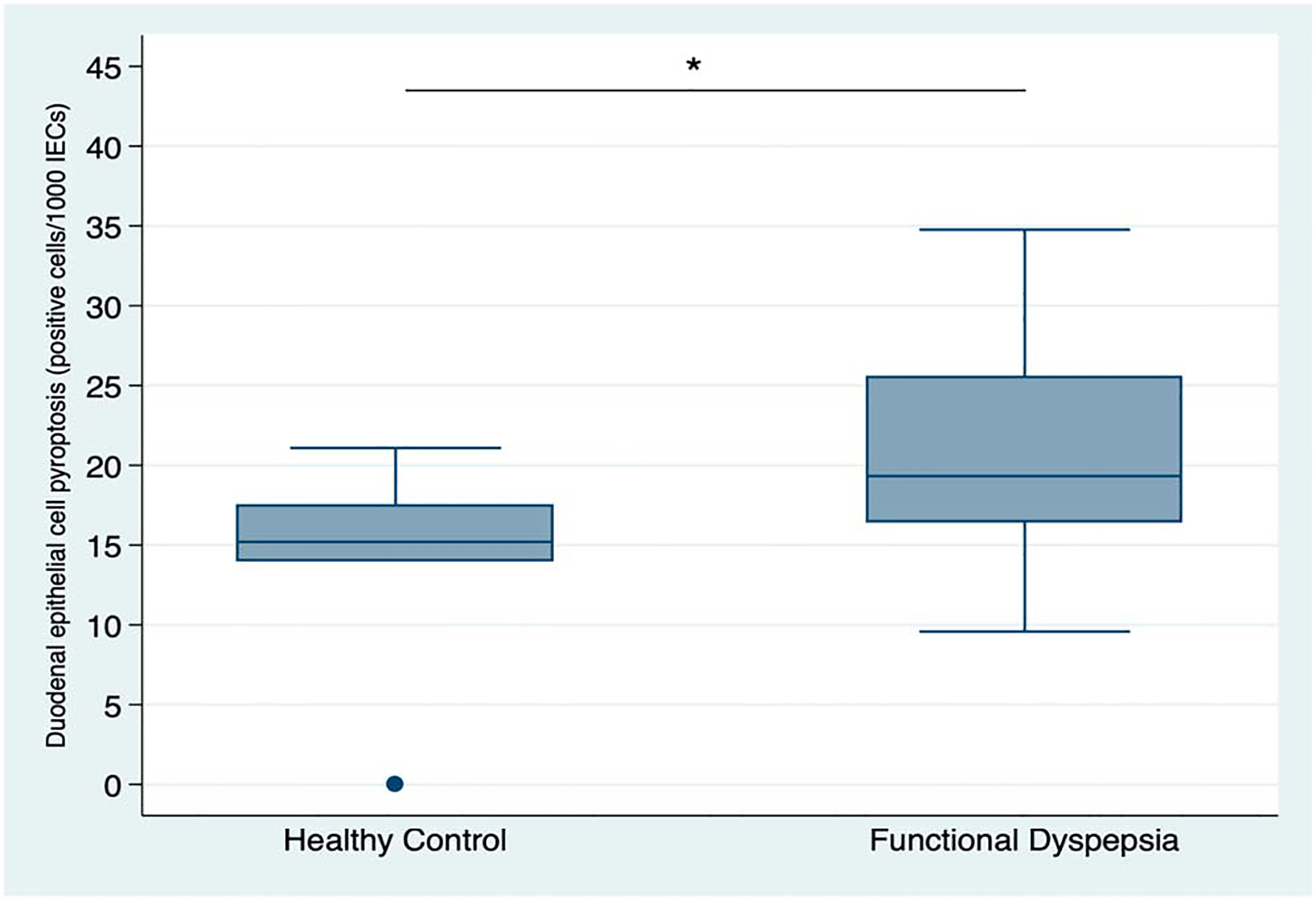
Quantitative assessment of duodenal epithelial cells staining positive for activated caspase-1 (immunohistochemistry) on biopsy samples from healthy controls (n = 6) and patients with FD(n = 14). *P = 0.04.

Quantitative assessment of duodenal epithelial cells staining positive for activated caspase-1 (immunohistochemistry) on biopsy samples from healthy controls (n = 6) and patients with FD(n = 14). *P = 0.04.