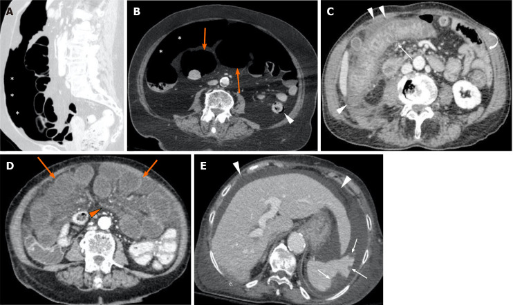
Figure 1.
Computed tomography images from positive coronavirus disease 19 patients with abdominal signs and symptoms. A and B: The computed tomography (CT) scan revealed pneumoperitoneum (asterisks, A and B), dilated bowel loops with thin non-enhancing walls (orange arrows, B) and focal areas of pneumatosis in the descending colon (arrowhead, B), in keeping with intestinal ischemia. The main mesenteric vessels were patent (not shown); C: CT evidence of ischemic colitis involving the transverse and right portions of the colon: Markedly thickened and layered walls (thin arrow), compared to other normal appearing small and large bowel loops (curved arrow), with associated free fluid and oedematous stranding of the adjacent fat tissue (arrowheads); D and E: Axial CT scan shows multiple small bowel loops moderately dilated, with reduced thickness and poorly enhancing walls (orange arrows, D); concomitant diffuse oedema of the mesentery (orange arrowhead, D). In the same patient there were associated multiple peripheral splenic infarcts (withe arrows, E) with areas of mottled increased attenuation; the main splenic vessels were patent (not shown). Diffuse ascites (white arrowheads, E) is noted.

