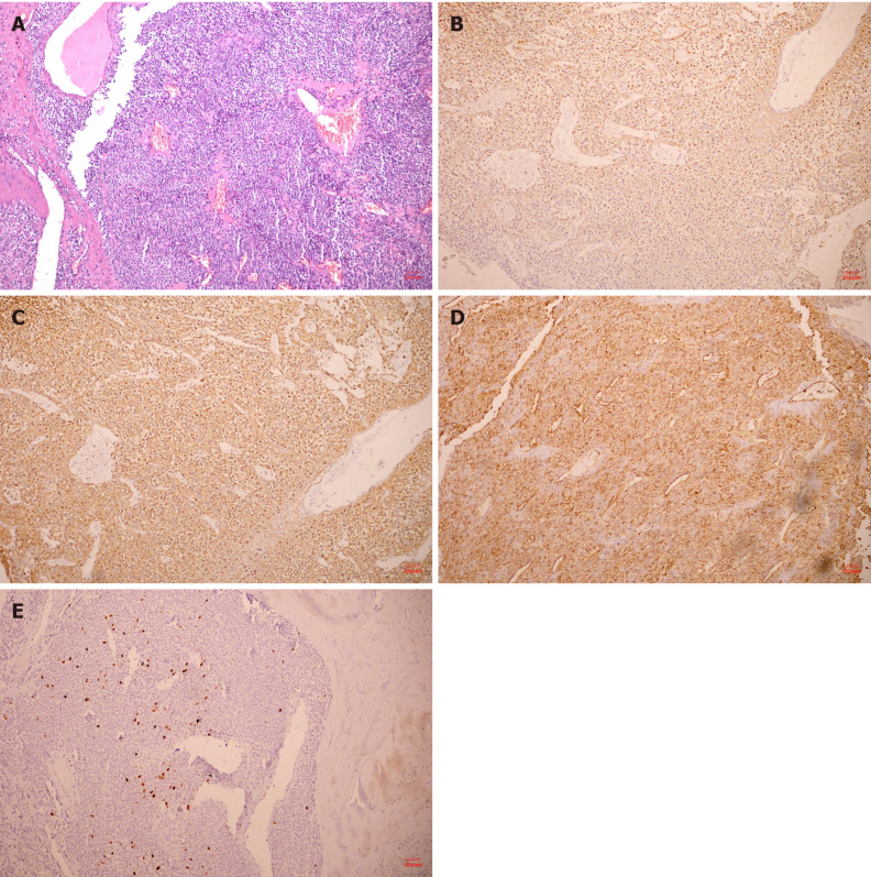
Figure 4.
Hematoxylin and eosin pathological sections and immunohistochemistry after operation. A: The tumor was composed of aggregated round and fusiform cells surrounded by capillaries (10 ×); B: Immunohistochemistry showed spinal muscular atrophy (+, 10 ×); C: H-caldesmon (+, 10 ×); D: cluster of differentiation 34 (+, 10 ×); E: A Ki-67 positive rate of 2% (10 ×).

