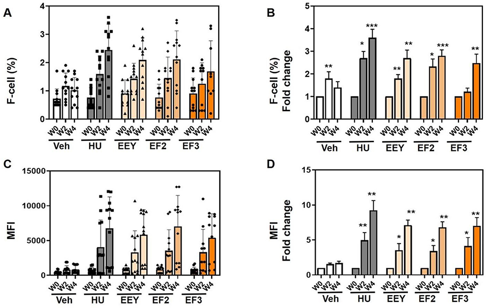
Fig. 3. Benserazide increases HbF in young β-YAC mice.
β-YAC mice (6–8 weeks old) were treated with water (Veh) and hydroxyurea (HU) controls and the racemic (R,S)-benserazide HCL (EEY), (R)-benserazide HCL (EF2) and (S)-benserazide HCL (EF3) drug formulations. Three treatment replicates (n=12) were combined and analyzed as described and blood collected for flow cytometry analysis (See Methods). A) Shown are raw data for the F-cells percent (%) at week 0 (W0), week 2 (W2) and week 4 (W4). B) Shown is the fold change in F-cells over same time-period. C) To quantify mean fluorescence intensity (MFI) was quantified. Shown are the MFI mean over same time-period. D) Shown are fold changes in MFI. Statistical analysis as described in Fig. 1.

