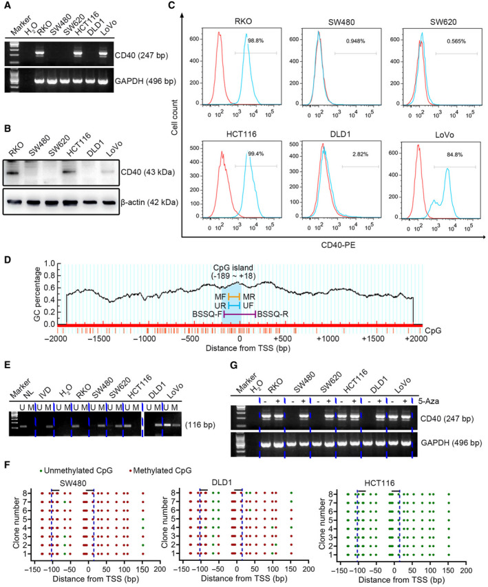
Fig. 7.

The expression of CD40 is regulated by promoter methylation in CRC cell lines. (A) mRNA (247 bp), (B) total protein (43 kDa) and (C) membrane expression of CD40 in six CRC cell lines (RKO, SW480, SW620, HCT116, DLD1 and LoVo). GAPDH (496 bp) and β‐actin (42 kDa) were used as the loading controls. (D) Schematic diagram of a CpG island in the promoter region of CD40. (E) Methylation status of CD40 (116 bp) detected by MSP in CRC cell lines. (F) BSSQ of CD40 performed in SW480, DLD1 and HCT116 cell lines. Red solid dots represent methylated CpG sites, and green solid dots denote unmethylated CpG sites. The horizontal black bar demarcates the primers of MSP, which are included in the region of BSSQ. (G) mRNA expression of CD40 (247 bp) with (+) or without (−) treatment of 5‐Aza. GAPDH (496 bp) was used as the loading control. BSSQ‐F, bisulfite sequencing forward primer; BSSQ‐R, bisulfite sequencing reverse primer; IVD, in vitro methylated DNA; M, methylated alleles; MF, methylation forward primer; MR, methylation reverse primer; NL, normal lymphocyte DNA; UF, unmethylation forward primer; U, unmethylated alleles; UR, unmethylation reverse primer.
