
FIGURE 7.
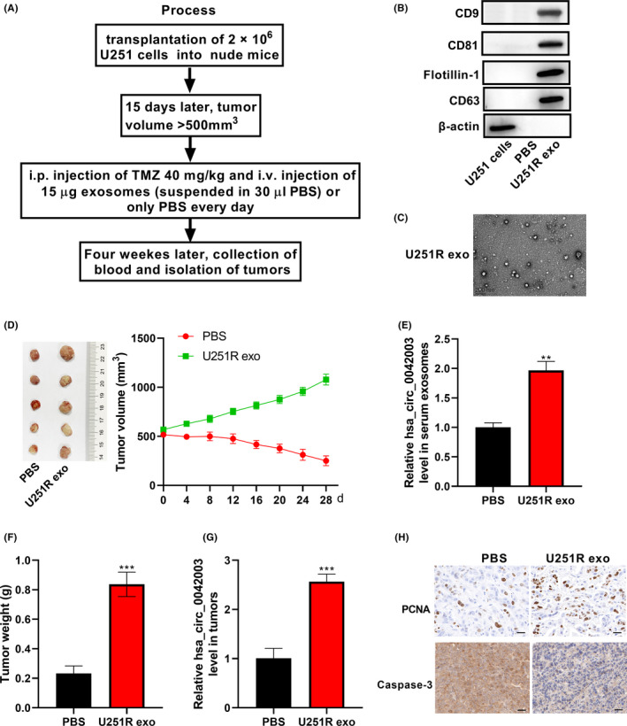
Exosomes derived from chemoresistant cells decrease the drug susceptibility of chemosensitive cells in vivo. A, Experimental design diagram of the xenograft model. Exosomes derived from U251R cells were collected. B, Expression of exosome (exo) marker proteins flotillin‐1, CD63, CD9, and CD81 was analyzed using western blot assay. C, Morphology of exosomes was observed using transmission electron microscopy. D, Volume of xenograft tumors changed during TMZ and exosome treatment. After 28 days of treatment, mouse serum was collected and serum exosomes were extracted. E, Quantitative real‐time PCR (qRT‐PCR) was used to assess the level of hsa_circ_0042003 in serum exosomes. F, Tumors were resected from mice at the same time on day 28 of treatment, and the tumor weight is shown. G, Level of hsa_circ_0042003 in xenograft tumors was analyzed using qRT‐PCR. H, Immunohistochemical analysis of the expression of proliferating cell nuclear antigen (PCNA) and caspase‐3 in xenograft tumors. Scale bar = 20 μM. **P < .01; ***P < .001